Percutaneous coronary Intervention (PCI): PCI is a technique with which blocks can be removed and thus it can be used to clear blocks or even cure acute heart attacks. A special catheter is used by the Interventional Cardiologists through a needle-entry in the hand. The cardiologist gets access to the blocked artery and crushes the blocks. This result in restoration blood supplies to the heart and thus prevents muscle damage. The procedure of PCI saves lives, and lowers the risks of complications and long term mortality for a large group of Indian patients.
EVAR/TEVAR: Endovascular Aneurysm Repair is a procedure used to treat aneurysms. Aneurysms are anomalous bulge of the blood vessel. Aneurysms usually take place in the arteries supplying to the abdominal area.
Here I must inform the readers that there are times this method is administered in the thoracic region; it’s called Thoracic Endovascular Aneurysm Repair (TEVAR). While conducting this particular procedure, a stretched-out stent is positioned in the aorta with the aid of a catheter, without any invasive surgeries.
TAVR: The technique known as the Transcatheter Aortic Valve Replacement is usually administered in order to substitute a significantly important valve in the heart. The Aortic valve controls the flow of blood into the heart. As age advances, the aortic valve may become thick and narrow. Additionally, the valve may develop calcification, and the condition could lead to death. To counter that, this process is used to put a replacement valve in an attempt to substitute for the damaged existing aortic valve, using a catheter without any surgery.
Rotablation Angioplasty: In Rotablation Angioplasty, calcified deposits in the artery are removed. The technique uses a catheter attached to a tiny drill-like motor and it is introduced into the artery.
The machine then drills away the strong, calcified deposits without damaging the artery. After the Rotablation technique is done, a balloon is sent in with a stent to keep the artery from collapsing.
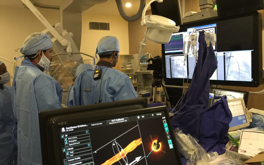
Furthermore, cathlabs are also utilized to get a good look into the blood vessel in the heart. This uses highly advanced imaging techniques like the Intravascular Ultrasound, Optical Coherence Tomography and others.
Procedures like these are extremely advanced. Moreover, they give absolutely clear and more high-definition images of the blood vessels. These methods give more vital additional information than the traditional angiogram.
I and my team have been aided a lot by techniques such as Fractional Flow Reserve, to take precise and trustworthy measurements of blood flow into the heart. Most significantly, FFR also helps us to understand the scale and scope of the blocks in the blood vessels and need for stents or not.
IVUS and OCT when combined with FFR, the set forms an extremely beneficial option which can tell the doctors if a complex surgery is required, or whether simple medication is enough to remove the blocks. This will obviously help in cost-savings, and provide a better quality of life for patients.