With the advancement of medical science and medical technology, our imagination was absolutely at a setback around 15 years back. And I have keenly felt this every day and mostly when I am inside the cathlab of Cardiology in a hospital like the Apollo Hospitals in Chennai.
Rene Laennec had invented the stethoscope and since its initiation several other physicians have tried to refine the device. In fact, the latest version of stethoscope that we use was designed and finalised by George Philip Cammann. Most importantly, it is considered as the significant medical invention. This is because it was the first invention which permitted a doctor to look into a patient’s body without surgically opening the body. While the stethoscope aided the doctors to hear the sounds created by the heart and lung of a patient, physicians utilize the device as a guide to diagnose diseases.
This clearly indicates that, since then, diseases were seen not as an assortment of symptoms, but as a chief cause, giving out a certain number of symptoms.
To be exact, medical experts have tried to reach the goal where medical science and technology would be used to refine this process, that is, perfectly locating the cause of the problem, and confirm the best way to treat it.
We have previously discussed that Fractional Flow Reserve (FFR)is one of the tools which are used in the cardiologist’s kit. Now, let’s talk about the next big thing in cardiology – Intra Coronary Imaging. In my opinion, this process is like taking a photograph inside the blood vessels.
Imaging of this sort is mostly done through two processes. The first one is based on sound and it is called Intravascular Ultrasound (IVUS). It is basically the same technology which is used to do ultrasound scan (e.g.-abdomen)
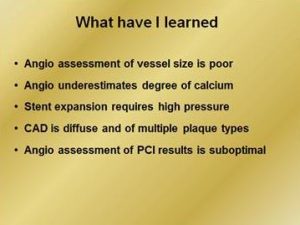
I believe angiogram to be just a luminogram and not an actual representation of what’s going on inside. I can undoubtedly note that angiogram shows the locations as the dye goes through. However, the test cannot show the accurate dimensions of the arteries.


It has been 30 years that IVUS is being administered and a massive amount of research provides us with evidence for its efficiency. The process involves the placing of a miniaturised probe inside the artery, and a scan is done. After that, the probe gives out high-frequency sound waves which are analysed by a particular computer system. Based on the reflected sound waves produces images which gives information on the inner wall of the artery, the width of any plaque, and finally the lumen or obtainable space for the flow of blood.
The biggest advantage IVUS has over traditional angiogram is that it informs the doctor about the presence and nature of the block, its thickness and whether it is weakening the artery. In actuality, an angiogram may give a picture of the block in one part of the blood vessel. However, the angiogram doesn’t give an image of whether the entire circumference of the vessel has important plaque build-up and over a large section. During a scenario like this – IVUS is of the most noteworthy procedure.
As I have understood,for beginners IVUS possesses a greater learning curve. Most significantly, the test is extremely valuable to recognize the actual dimensions of the blood vessel.
Through the use of IVUS it can be determined that the plaque build-up in the artery will massively affect the thickness and strength of the artery wall. It can be noted that if the build-up is heavy and the artery is weak – it means that the artery will not be capable to sustain the pressure, and may burst.


